The Diagram Represents A Process Used To Modify Bacterial Ce
Why gram positive bacteria are purple in color after using safranin How to draw a bacteria easy/bacteria drawing Restriction enzymes
Bacteria Cell Structure - Prokaryotic Cell Diagram - 1258x1024 PNG
Gram procedure staining stain positive bacteria microbiology purple color results principle steps after why bacterial method safranin classification diagram using Draw a labelled diagram of a bacterial cell. Enzymes plasmid restriction bacteria dna biology enzyme function bacterial rna
Gram procedure staining stain positive bacteria microbiology results principle color purple steps method bacterial why after classification protocol step diagram
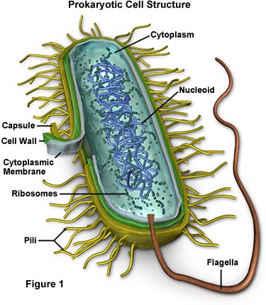
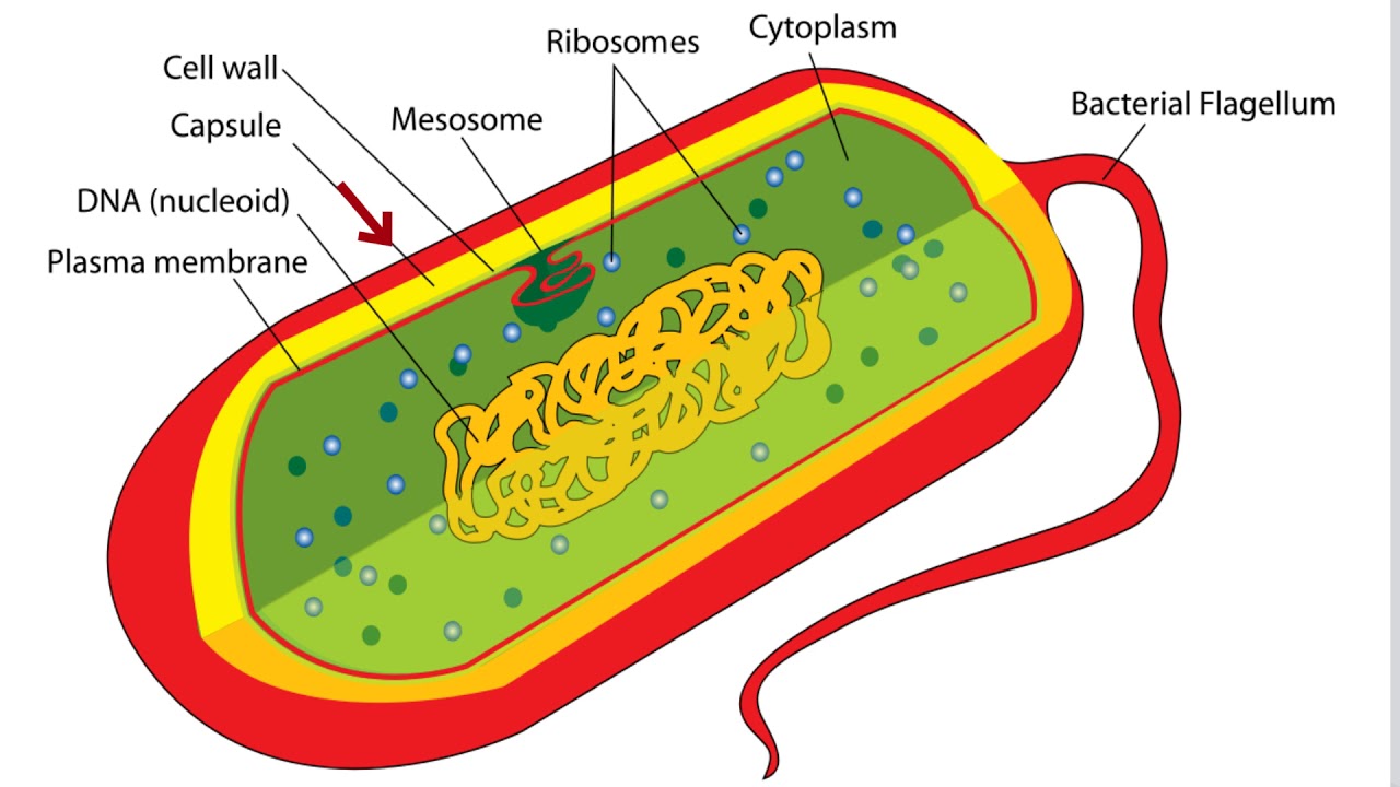
Cell structure cells bacteria bacterial prokaryotic diagram biology cellular organelles functions like model ribosomes dna nucleus which found prokaryote largeFig. 1 the schematic diagram of bacterial cell structure. in 2023 Solved move the images of bacterial cells into the chart toProkaryotic structure prokaryotes bacteria bacterial labeled membrane organelles eukaryotic prokaryote nucleus functions found mitochondria plasma within.
Gram staining: principle, procedure, results • microbe onlineBacterial cell diagram and functions Bacteria cell structure3.3 unique characteristics of prokaryotic cells – microbiology.

Bacteria cell structure
Bacterial structuresBacterial cell structure blank Bacteria- definition, diagram and classificationMolecular expressions cell biology: bacteria cell structure.
Difference between prokaryotic and eukaryotic cellGram procedure staining stain positive bacteria microbiology results purple principle color steps bacterial method after why classification diagram safranin cell Cells bacterial chart gram solved move into negative positive illustrate step color answer problem been has stain process eachSolved move the images of bacterial cells into the chart to.

Gram bacteria microscope procedure staining negative observing rsscience microbiology principle experiment difference
Cells: bacterial diagramCell prokaryotic bacteria prokaryotes bacterial nucleus primitive organisms microorganisms distinct lack because neat Cell bacterial anatomy ribosomes nucleoid pili bacillus vector flagellum labeling vecteezyBacterial cell anatomy in flat style. vector modern illustration.
Solved move the images of bacterial cells into the chart toProkaryotic bacteria prokaryotes microbiology membrane plasma ecampusontario pressbooks pub structure Bacteria cell structure17+ bacteria labelled diagram.

17+ bacteria labelled diagram
Bacterial cell labelled diagram| how to draw bacteria cell 🦠Inside 107 and 109 Science laboratory medical gram negative bacteria cell wall microbiologyCell prokaryotic prokaryotes bacteria membrane bacterial labeled organelles prokaryote eukaryotic nucleus lack classconnection mitochondria chessmuseum eukaryotes.
Bacterial structuresIgcse diagram Solved earch the web welcome saDiagram of igcse biology diagrams.
Prokaryotes are the organisms which have primitive nucleus. all
Bacterial bacteria salmonella necrotizing fasciitis biology estructura microorganisms organism micro sketchite celulas membrane microbiology parashuramBacteria cell structure Observing bacteria under the microscope17+ simple diagram of bacteria.
[solved] draw and label a typical bacterial cell, then provideBacteria cell structure Pin on labs.


![[Solved] Draw and label a typical bacterial cell, then provide](https://i2.wp.com/www.coursehero.com/qa/attachment/12940603/)
